 Asclepio
Inside a tooth in 3D
A tooth has four tissues: enamel, dentin, cementum and pulp. The crown, the visible part, is covered with enamel; the root is covered with cementum and…
Explained for every age
For kids (7–11)
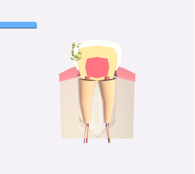
Here you see a molar cut in half. On the outside is the enamel, hard and shiny; underneath, the dentin; and in the center, the pulp, with nerves and blood vessels. Watch how plaque causes tooth decay… and how the toothbrush sweeps it away!
For teens (12–16)
A tooth has four tissues: enamel, dentin, cementum and pulp. The crown, the visible part, is covered with enamel; the root is covered with cementum and set into the bone. Around it, the periodontal ligament, the alveolar bone and the gum hold it in place. In the scene, dental plaque grows, its acid opens a cavity that reaches the dentin, and then the toothbrush cleans off the plaque; the cavity that has already formed doesn't heal on its own.
Pre-Med (17+)
Cross-section of a molar with its four tissues: enamel, dentin, cementum and pulp. The anatomical crown is the enamel-covered area above the cementoenamel junction (neck); the root, below it, is covered with cementum, and both are made mostly of dentin. The periodontium (cementum, periodontal ligament, alveolar bone and gingiva) anchors the tooth and allows touch and pressure to be sensed. Vessels and nerves enter the pulp through an opening at the apex of the root. The molar in the scene has two roots, as lower molars usually do (upper molars usually have three). Simplifications: the bacteria are exaggerated so they can be seen, and the cementum is not shown as a separate layer.
What you can explore
EnamelDentinDental pulpPeriodontal ligamentAlveolar boneGingiva (section)Dental nerve and vesselsDental plaqueDental cariesToothbrushing
Missions
- Save the tooth! Explorer
- Layer by layer Discoverer
- Dentin-pulp complex and periodontium Pre-Med