 Asclepio
Real cell (electron microscope) in 3D
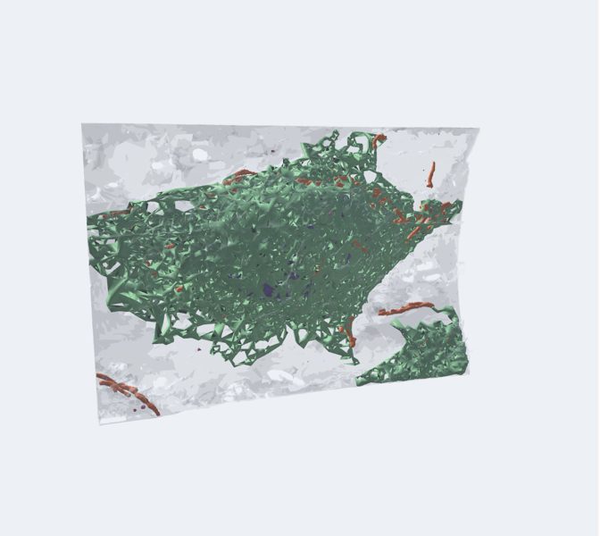
A HeLa cell (a human cell line used in laboratories around the world since 1951) reconstructed by FIB-SEM electron microscopy at 4 nm resolution and…
Explained for every age
For kids (7–11)
This is not a drawing: it is a real cell, photographed layer by layer with an electron microscope and rebuilt in 3D by scientists at the Janelia institute. Look how tightly packed everything is inside: the mitochondria look like worms and the reticulum is a huge network. It is flat like a pancake because it grew stuck to the bottom of a lab dish.
For teens (12–16)
A HeLa cell (a human cell line used in laboratories around the world since 1951) reconstructed by FIB-SEM electron microscopy at 4 nm resolution and segmented by artificial intelligence in the OpenOrganelle project. Each organelle has the real shape it had: branched mitochondria, a reticulum that fills the cytoplasm, the Golgi stacked next to the nucleus. The cell is flat because it grew attached to a dish; you see it as under the microscope, from above. Use the cut to see inside. The straight edges are not part of the cell: they are the boundary of the block the microscope scanned, which cut off part of it.
Pre-Med (17+)
FIB-SEM volume at 4 × 4 × 5.24 nm of a HeLa cell (jrc_hela-2), organelle segmentation by neural networks (COSEM, Heinrich et al. 2021). Real meshes of the plasma membrane, nucleus, ER, Golgi, mitochondria, endosomes/lysosomes and vesicles. HeLa: cervical adenocarcinoma of Henrietta Lacks (1951), HPV-18 positive, hypertriploid; immortal reference cell line. Note the networked mitochondrial morphology and the ER cisternae compared with textbook figures.
Missions
- A real cell Explorer
- Real organelles Discoverer
- Ultrastructure and ethics Pre-Med